
AI-Powered Pathology, Redefining Precision Diagnostics
First-to-market certified AI solutions transforming Atherosclerosis, Liver, Kidney and Brain disease diagnostics
AI-POWERED HISTOPATHOLOGY PLATFORM

HistoSuite
Our comprehensive portfolio of AI-based products purpose-built for preclinical histopathology assessment. HistoSuite transforms routine stained whole-slide images into rich, structured datasets elevating efficacy analysis, and strengthening translational research.
Quantified efficacy endpoints
Precise, quantitative measurements of lesion area, composition, and morphological features across entire tissue sections.
Standardized & reproducible assessments
Eliminate inter- and intra-reader variability with fully automated AI analysis delivering identical results regardless of operator.
Multi-omics integration
Combine AI-derived histopathological data with transcriptomics,
proteomics, and biomarker datasets for comprehensive multimodal insights.
AI MASH HISTOPATHOLOGY: Liver AI
TNO's AI histopathology turns liver tissue samples into objective, reproducible MASH and fibrosis read-outs. Scoring steatosis, inflammation and fibrosis similar to pathologists, but across 100% of the tissue and in a fraction of the time.
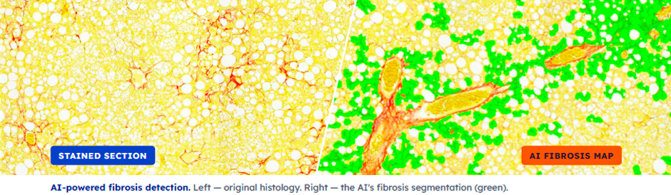
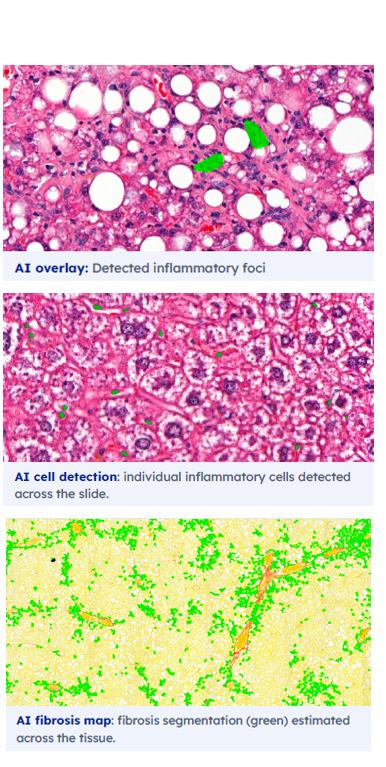
Quantify liver histology faster and more accurate with AI
Our AI reads whole-slide images of liver tissue and reproduces the pathologist's reasoning. The result is standardized, repeatable quantification that removes inter- and intra-reader variability and scales to hundreds of slides. It is built for preclinical studies, where scoring large cohorts consistently is hard to achieve by manual review.
Three key MASH markers, Quantified by AI
Inflammation
The AI finds and counts inflammatory-cell aggregates much as a pathologist does and reports their density per mm2, estimated across 100% of the tissue (a pathologist samples ~30%). Because individual cells are detected, it also reports cells per aggregate and total counts, statistics infeasible to manually collect at scale.
Fibrosis
It estimates the percentage of tissue affected by fibrosis the way a pathologist does, not as an abstract pixel-level score. The AI scores closely track expert assessments across study groups.
Steatosis
Macro- and microvesicular steatosis are quantified as affected-tissue estimates, reproducing the pathologist's read in an automated, reproducible way. Additional markers are on the roadmap.
CASE STUDY ・ LIVER FIBROSIS
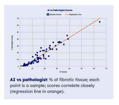 AI fibrosis scores track the pathologist
We tested whether AI fibrosis read-outs can capture early treatment effects. Mice on a 28-week high-fat diet (HFD) were then kept on HFD, switched to a lean chow diet, or given a long-acting FGF21 analogue - a direct anti-fibrotic - for 4 or 8 weeks, with a start-of-treatment group as reference.
Both pathologist and AI analyses show HFD increases fibrosis, while the dietary switch and FGF21 inhibit its development. Across every group, the AI's scores correlate closely with the pathologist's.
AI BRAIN PATHOLOGY : Brain AI
TNO's AI histopathology algorithms turns brain sections into objective, region-level read-outs of neurodegeneration and neuroinflammation.
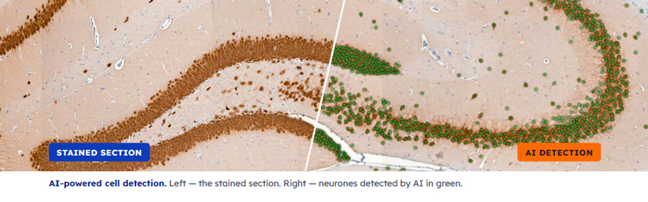
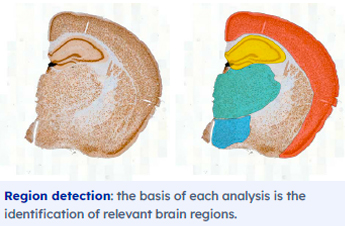
Quantify brain sections faster and more accurately with AI
Our AI reads whole-slide images of stained brain sections, segments the brain regions, and then detects individual cells. The result is a reproducible quantification that removes reader variability and scales to large preclinical cohorts.
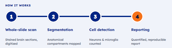 Multi marker readouts
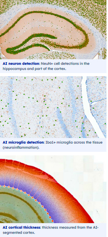
Neuron detection and density (NeuN)
Every NeuN+ neuron is detected and counted within its region, giving objective neuronal-density and neuron-loss read-outs. With more than 30k neurons per slice, such a measurement would be infeasible by hand.
Microglial activation (Iba1)
Iba1+ microglia are detected across the section to quantify microglial activation and the regional spread of neuroinflammation.
Cortical thickness
The AI-segmented cortex is measured directly into reproducible cortical-thickness maps across the whole section, a structural read-out infeasible to produce by hand at scale.
Why it matters
● Faster and cost-saving: shorter turnaround than manual scoring of large cohorts.
● Replicates the pathologist: counts cells and estimates affected tissue the way an expert scores it.
● Reads 100% of the section: densities and areas computed over the whole tissue, not a sampled subset.
● Markers beyond manual review: per-region cell counts and densities that are infeasible by hand.
● Standardized and reproducible: removes inter- and intra-reader variability.
AI-Powered Preclinical Atherosclerosis Research
The APOE3-Leiden (E3L) and APOE3-Leiden.huCETP (E3L.CETP) mouse models are well-established, translational models for evaluating therapies targeting plasma lipids, lipoprotein metabolism, and atherosclerosis, with proven responsiveness to clinically used lipid-lowering drugs in both preventive and therapeutic study designs. AI-assisted assessment enhances these studies by standardizing lesion quantification, reducing inter- and intra-reader variability, and improving sensitivity to detect treatment effects. TNO Athero AI transforms routine H&E-stained aortic root sections into objective, quantitative datasets that strengthen confidence in your efficacy data.
 Score with consistency
Increase scoring confidence by minimizing variability in atherosclerosis trials and research. TNO Athero AI ensures that every lesion is evaluated against the same objective criteria, eliminating the subjective differences that arise between human readers and across different reading sessions.
Segment and classify
TNO Athero AI accurately segments plaques and characterizes their composition, including necrotic core, calcification, fibrous tissue, and smooth muscle content. This detailed compositional analysis reveals treatment effects that conventional area-based measurements would miss entirely.
Efficient workflows
Speed up trials by reducing turnaround times through AI-assisted reading. Automated analysis processes hundreds of whole-slide images in a fraction of the time required for manual pathologist review, enabling faster data delivery without compromising quality or detail.
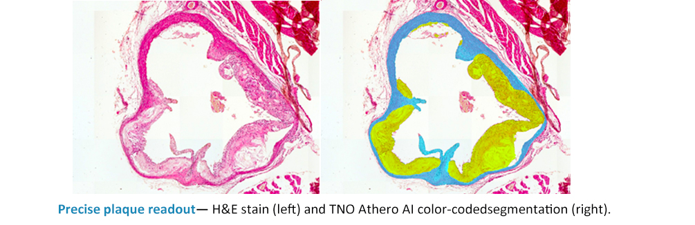
Precise and standardized plaque readout
TNO Athero AI segmentation enables precise and standardized plaque readout, accurately delineating lesion boundaries across the complete aortic root cross-section. Automated analysis reduces turnaround time and minimizes reader variability, while clear, color-coded outputs make results straightforward to interpret in atherosclerosis research and preclinical efficacy studies. Each plaque component is mapped with pixel-level precision, providing quantitative measurements that go far beyond what manual assessment can achieve. AI segmentation accurately delineates lesion boundaries across the complete aortic root cross-section, mapping each plaque component with pixel-level precision. Small, early-stage lesions are consistently detected.
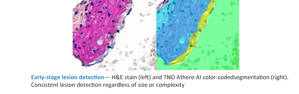
Consistent detection of early-stage lesions
TNO Athero AI analysis helps ensure that even small, early-stage atherosclerotic lesions are consistently detected and accurately delineated across all study samples. In preventive study designs where lesion burden may be significantly reduced by effective treatment, this sensitivity is critical. The AI model reduces the risk of overlooked features and strengthens the reliability of lesion assessment within and across studies, ensuring that subtle but meaningful treatment effects are captured with statistical confidence.
How HistoSuite Works
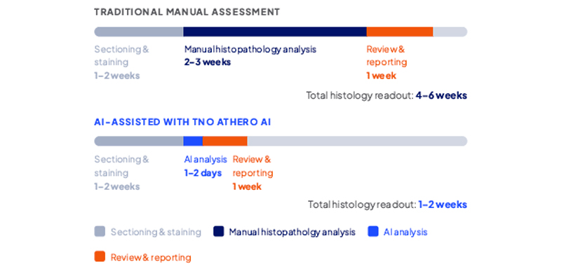 By automating the most labor-intensive step in plaque assessment, TNOAthero AI compresses analysis windows from weeks to days ? without sacrificing depth, accuracy, or reproducibility.
TNO Kidney AI: under construction
